Researchers have developed ultra-flexible brain probes that accurately record brain activity without causing tissue damage.
This opens up new avenues for the treatment of a range of neurological and neuropsychiatric disorders.
Neurostimulators, also known as brain pacemakers, send electrical impulses to specific areas of the brain via special electrodes. It is estimated that some 200,000 people worldwide are now benefiting from this technology, including those who suffer from Parkinson’s disease or from pathological muscle spasms.
According to Mehmet Fatih Yanik, professor of neurotechnology at ETH Zurich, further research will greatly expand the potential applications: instead of using them exclusively to stimulate the brain, the electrodes can also be used to precisely record brain activity and analyze it for anomalies associated with neurological or psychiatric disorders.
In a second step, it would be conceivable in future to treat these anomalies and disorders using electrical impulses.
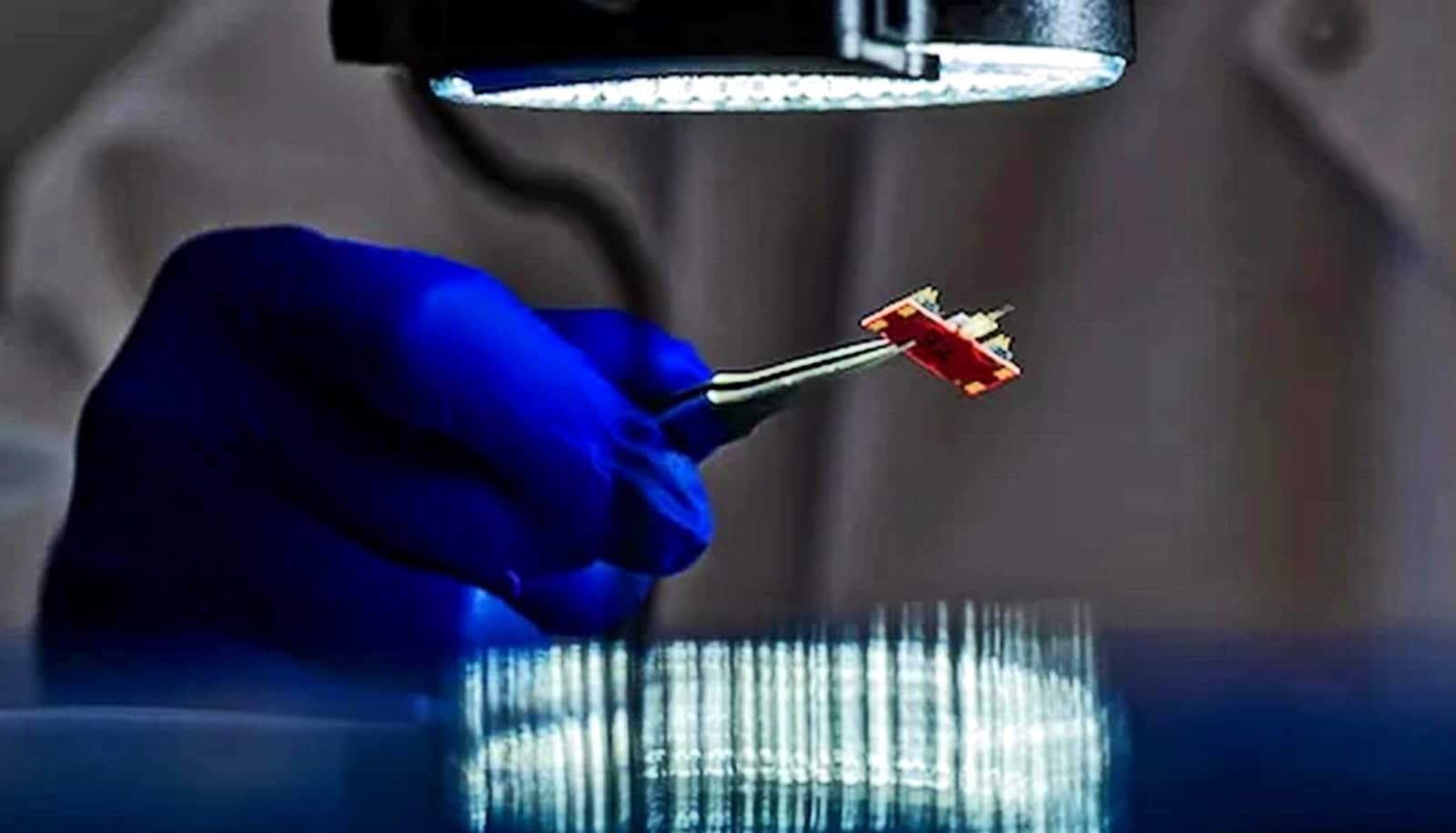
To this end, Yanik and his team have now developed a new type of electrode that enables more detailed and more precise recordings of brain activity over an extended period of time. These electrodes are made of bundles of extremely fine and flexible fibers of electrically conductive gold encapsulated in a polymer.
Thanks to a process developed by the ETH Zurich researchers, these bundles can be inserted into the brain very slowly, which is why they do not cause any detectable damage to brain tissue.
This sets the new electrodes apart from rival technologies. Of these, perhaps the best known in the public sphere is the one from Neuralink, an Elon Musk company. In all such systems, including Neuralink’s, the electrodes are considerably wider.
“The wider the probe, even if it is flexible, the greater the risk of damage to brain tissue,” Yanik explains. “Our electrodes are so fine that they can be threaded past the long processes that extend from the nerve cells in the brain. They are only around as thick as the nerve-cell processes themselves.”
The research team tested the new electrodes on the brains of rats using four bundles, each made up of 64 fibers. In principle, as Yanik explains, up to several hundred electrode fibers could be used to investigate the activity of an even greater number of brain cells. In the study, the electrodes were connected to a small recording device attached to the head of each rat, thereby enabling them to move freely.
In the experiments, the research team was able to confirm that the probes are biocompatible and that they do not influence brain function. Because the electrodes are very close to the nerve cells, the signal quality is very good compared to other methods.
At the same time, the probes are suitable for long-term monitoring activities, with researchers recording signals from the same cells in the brains of animals for the entire duration of a ten-month experiment. Examinations showed that no brain-tissue damage occurred during this time.
A further advantage is that the bundles can branch out in different directions, meaning that they can reach multiple brain areas.
In the study, the researcher used the new electrodes to track and analyze nerve-cell activity in various areas of the brains of rats over a period of several months. They were able to determine that nerve cells in different regions were “co-activated”. Scientists believe that this large-scale, synchronous interaction of brain cells plays a key role in the processing of complex information and memory formation.
“The technology is of high interest for basic research that investigates these functions and their impairments in neurological and psychiatric disorders,” Yanik explains.
The group has teamed up with fellow researchers at the University College London in order to test diagnostic use of the new electrodes in the human brain. Specifically, the project involves epilepsy sufferers who do not respond to drug therapy. In such cases, neurosurgeons may remove a small part of the brain where the seizures originate. The idea is to use the group’s method to precisely localize the affected area of the brain prior to tissue removal.
There are also plans to use the new electrodes to stimulate brain cells in humans.
“This could aid the development of more effective therapies for people with neurological and psychiatric disorders,” says Yanik.
In disorders such as depression, schizophrenia, or OCD, there are often impairments in specific regions of the brain, which lead to problems in evaluation of information and decision making. Using the new electrodes, it might be possible to detect the pathological signals generated by the neural networks in the brain in advance, and then stimulate the brain in a way that would alleviate such disorders.
Yanik also thinks that this technology may give rise to brain-machine interfaces for people with brain injuries. In such cases, the electrodes might be used to read their intentions and thereby, for example, to control prosthetics or a voice-output system.
The research appears in Nature Communications.
Funding for this research came in part from a Consolidator Grant from the European Research Council (ERC) awarded to Mehmet Fatih Yanik in 2018 and the Swiss National Science Foundation’s Sinergia programme.
Source: ETH Zurich