Researchers have begun to solve one of COVID-19’s biggest and most life-threatening mysteries: how the virus causes “silent hypoxia,” a condition where oxygen levels in the body are abnormally low.
Those low oxygen levels can can irreparably damage vital organs if gone undetected for too long.
More than six months since COVID-19 began spreading in the US, scientists are still solving the many puzzling aspects of how the novel coronavirus attacks the lungs and other parts of the body.
Despite experiencing dangerously low levels of oxygen, many people infected with severe cases of COVID-19 sometimes show no symptoms of shortness of breath or difficulty breathing.
Hypoxia’s ability to quietly inflict damage is why health experts call it “silent.” In coronavirus patients, researchers think the infection first damages the lungs, rendering parts of them incapable of functioning properly. Those tissues lose oxygen and stop working, no longer infusing the blood stream with oxygen, causing silent hypoxia. But exactly how that domino effect occurs has not been clear until now.
“We didn’t know [how this] was physiologically possible,” says Bela Suki, professor of biomedical engineering and of materials science and engineering at Boston University and one of the coauthors of the study in Nature Communications.
Some coronavirus patients have experienced what some experts have described as levels of blood oxygen that are “incompatible with life.” Disturbingly, Suki says that many of these patients showed little to no signs of abnormalities when they underwent lung scans.
To help get to the bottom of what causes silent hypoxia, biomedical engineers used computer modeling to test out three different scenarios that help explain how and why the lungs stop providing oxygen to the bloodstream.
They found that silent hypoxia is likely caused by a combination of biological mechanisms that may occur simultaneously in the lungs of COVID-19 patients, says lead author Jacob Herrmann, a biomedical engineer and research postdoctoral associate in Suki’s lab.
How healthy lungs work
Normally, the lungs perform the life-sustaining duty of gas exchange, providing oxygen to every cell in the body as we breathe in and ridding us of carbon dioxide each time we exhale.
Healthy lungs keep the blood oxygenated at a level between 95 and 100%—if it dips below 92%, it’s a cause for concern and a doctor might decide to intervene with supplemental oxygen. (Early in the coronavirus pandemic, when clinicians first started sounding the alarm about silent hypoxia, oximeters flew off the shelves as many people, worried that they or their family members might have to recover from milder cases of coronavirus at home, wanted to be able to monitor their blood oxygen levels.)
The researchers first looked at how COVID-19 affects the lungs’ ability to regulate where blood is directed. Normally, if areas of the lung aren’t gathering much oxygen due to damage from infection, the blood vessels will constrict in those areas. This is actually a good thing that our lungs have evolved to do, because it forces blood to instead flow through lung tissue replete with oxygen, which is then circulated throughout the rest of the body.
But Herrmann says preliminary clinical data has suggested that the lungs of some COVID-19 patients had lost the ability of restricting blood flow to already damaged tissue and, in contrast, were potentially opening up those blood vessels even more—something that is hard to see or measure on a CT scan.
Using a computational lung model, Herrmann, Suki, and their team tested that theory, revealing that for blood oxygen levels to drop to the levels observed in COVID-19 patients, blood flow would indeed have to be much higher than normal in areas of the lungs that can no longer gather oxygen—contributing to low levels of oxygen throughout the entire body, they say.
Next, they looked at how blood clotting may affect blood flow in different regions of the lung. When the lining of blood vessels get inflamed from COVID-19 infection, tiny blood clots too small to be seen on medical scans can form inside the lungs. They found, using computer modeling of the lungs, that this could incite silent hypoxia, but alone it is likely not enough to cause oxygen levels to drop as low as the levels seen in patient data.
Silent hypoxia hides in lungs
Last, the researchers used their computer model to find out if COVID-19 interferes with the normal ratio of air-to-blood flow that the lungs need to function normally.
This type of mismatched air-to-blood flow ratio is something that happens in many respiratory illnesses such as with asthma patients, Suki says, and it can be a possible contributor to the severe, silent hypoxia that has been observed in COVID-19 patients.
The models suggest that for this to be a cause of silent hypoxia, the mismatch must be happening in parts of the lung that don’t appear injured or abnormal on lung scans.
Altogether, the findings suggest that a combination of all three factors are likely to be responsible for the severe cases of low oxygen in some COVID-19 patients.
By having a better understanding of these underlying mechanisms, and how the combinations could vary from patient to patient, clinicians can make more informed choices about treating patients using measures like ventilation and supplemental oxygen.
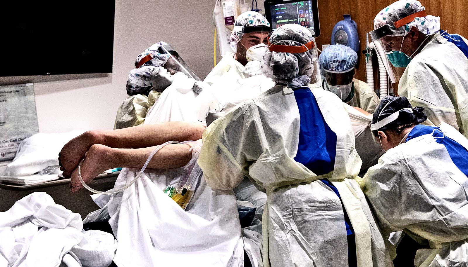
Researchers are currently studying a number of interventions, including a low-tech intervention called prone positioning that flips patients over onto their stomachs, allowing for the back part of the lungs to pull in more oxygen and evening out the mismatched air-to-blood ratio.
“Different people respond to this virus so differently,” Suki says. For clinicians, he says it’s critical to understand all the possible reasons why a patient’s blood oxygen might be low, so that they can decide on the proper form of treatment, including medications that could help constrict blood vessels, bust blood clots, or correct a mismatched air-to-blood flow ratio.
The National Heart, Lung, and Blood Institute supported the work.
Source: Boston University


