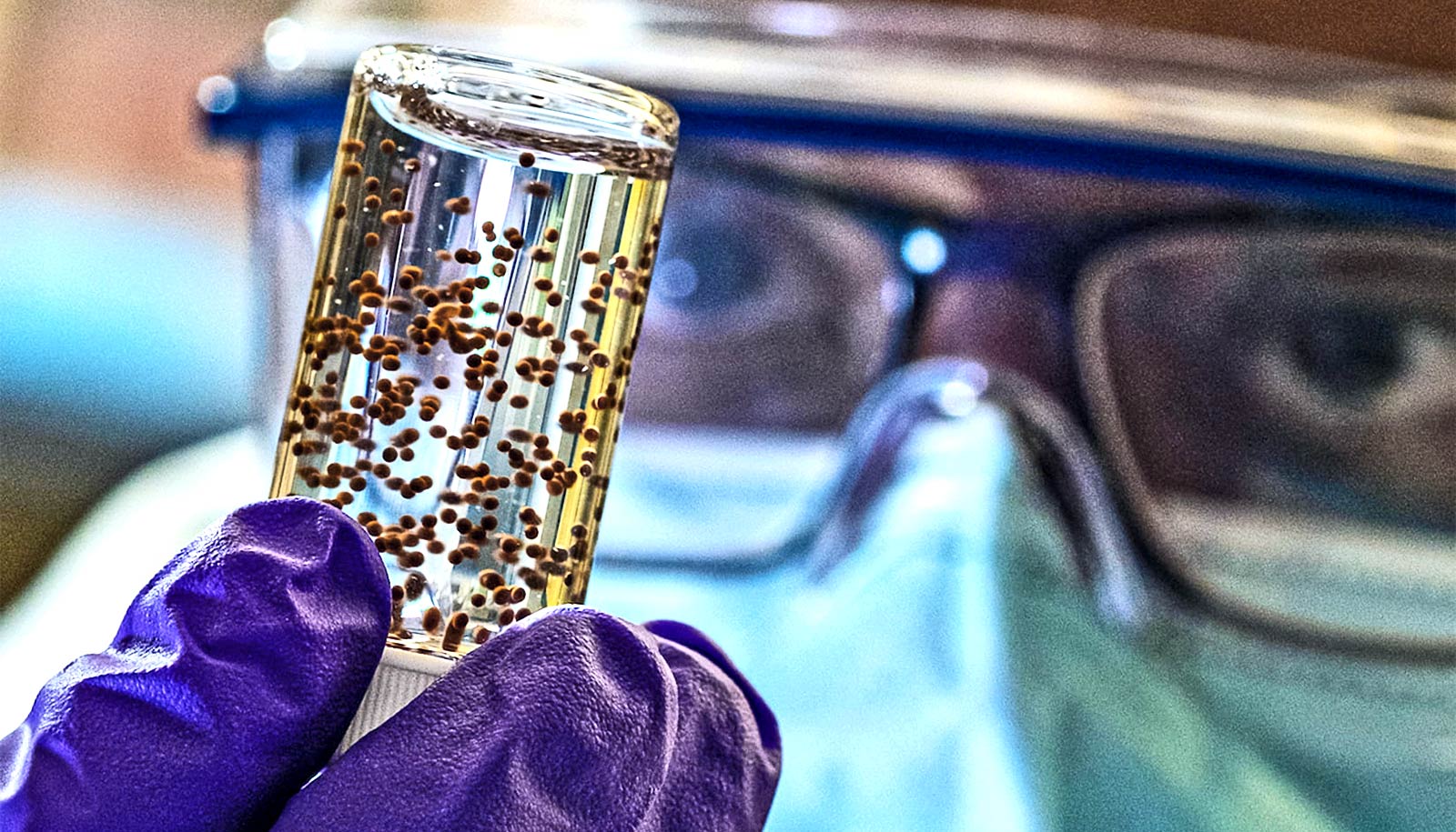
Shielding stem cells with a new biomaterial improves their ability to heal heart attack damage, a new study with rodents shows.
Researchers showed they could make capsules of wound-healing mesenchymal stem cells (MSCs) and implant them next to wounded hearts using minimally invasive techniques.
Within four weeks, they saw heart healing 2.5 times greater in animals treated with shielded stem cells than those treated with non-shielded stem cells.
Someone has a heart attack every 40 seconds in the United States. In each case, an artery that supplies blood to the heart becomes blocked and heart muscle tissue dies due to lack of blood. After a heart attack, hearts pump less efficiently and scar tissue from heart attack wounds can further reduce heart function.
“What we’re trying to do is produce enough wound-healing chemicals called reparative factors at these sites so that damaged tissue is repaired and restored, as healthy tissue, and dead tissue scars don’t form,” says Omid Veiseh, an assistant professor of bioengineering and CPRIT Scholar in Cancer Research at Rice University and co-lead author of the paper in the journal Biomaterials Science.
Stem cells and heart attacks
Prior studies have shown that MSCs, a type of adult stem cell produced in blood marrow, can promote tissue repair after a heart attack, says co-lead author Ravi Ghanta, associate professor of surgery at Baylor College of Medicine and a cardiothoracic surgeon at Harris Health’s Ben Taub Hospital. But in clinical trials of MSCs, “cell viability has been a consistent challenge,” he says.
“Many of the cells die after transplantation. Initially, researchers had hoped that stem cells would become heart cells, but that has not appeared to be the case. Rather, the cells release healing factors that enable repair and reduce the extent of the injury. By utilizing this shielded therapy approach, we aimed to improve this benefit by keeping them alive longer and in greater numbers.”
A few MSC lines have received approval for human use, but Veiseh says transplant rejection has contributed to their lack of viability in trials.
“They’re allogenic, meaning that they’re not from the same recipient,” he says. “The immune system perceives them as foreign. And so very rapidly, the immune system starts chewing at them and clearing them out.”
Veiseh has spent years developing encapsulation technologies specifically designed not to activate the body’s immune system. He cofounded Sigilon Therapeutics, a Cambridge, Massachusetts-based biotech company that is developing encapsulated cell therapeutics for chronic diseases. Trials of Sigilon’s treatment for hemophilia A are expected to enter the clinic later this year.
“The immune system doesn’t recognize our hydrogels as foreign, and doesn’t initiate a reaction against the hydrogel,” Veiseh says. “So we can load MSCs within these hydrogels, and the MSCs live well in the hydrogels. They also secrete the same reparative factors that they normally do, and because the hydrogels are porous, the wound-healing factors just diffuse out.”
In previous studies, Veiseh and colleagues have shown that similar capsules can keep insulin-producing islet cells alive and thriving in rodents for more than six months.
Delivering the capsules
In the heart study, co-lead author Samira Aghlara-Fotovat, a bioengineering graduate student in Veiseh’s lab, created 1.5-millimeter capsules that each contained about 30,000 MSCs. The researchers placed several of the capsules alongside wounded sections of heart muscle in animals that had experienced a heart attack.
The study compared rates of heart healing in animals treated with shielded and unshielded stem cells, as well as an untreated control group.
“We can deliver the capsules through a catheter port system, and that’s how we imagine they would be administered in a human patient,” Veiseh says.
“You could insert a catheter to the area outside of the heart and inject through the catheter using minimally invasive, image-guided techniques.”
The pericardium, a membrane that sheaths the heart, held the capsules in place, Veiseh says. Tests at two weeks showed that MSCs were alive and thriving inside the implanted spheres.
Looking ahead
More than 800,000 Americans have hearts attacks each year, and Ghanta is hopeful that encapsulated MSCs can one day treat some of them.
“With further development, this combination of biomaterials and stem cells could be useful in delivering reparative therapy to heart attack patients,” he says.
Veiseh says the pathway to regulatory approval could be streamlined as well.
“Clinical grade, allogenic MSCs are commercially available and are actively being used in patients for a range of applications,” he says.
Veiseh credits Aghlara-Fotovat with doing much of the work on the project.
“She basically executed the vision,” he says. “She developed the hydrogel formulation, the concept of how to package the MSCs within the hydrogel, and she did all the in vitro validation work to show that MSCs remained viable in the capsules.”
Ghanta comentors Aghlara-Fotovat, who worked in his lab at Baylor, including assisting in rodent surgeries and experiments.
“What attracted me to the project was the unmet clinical need in (heart attack) recovery,” Aghlara-Fotovat says. “Using hydrogels to deliver therapeutics was an exciting approach that aimed to overcome many challenges in the field of drug delivery. I also saw a clear path to translation into the clinic, which is the ultimate goal of my PhD.”
“I think one of the things that attracts students to my lab in particular is the opportunity to do translational work,” Veiseh says. “We work closely with physicians like Dr. Ghanta to address relevant problems to human health.”
Additional coauthors are from Rice and Baylor. An American Association of Thoracic Surgery Research Award; the Baylor College of Medicine Cardiovascular Research Institute; the Cancer Prevention Research Institute of Texas; the National Institutes of Health; a Rice University Academy Fellowship; the Emerson Collective; and the National Institutes of Health/National Heart, Lung and Blood Institute Research Training Program in Cardiovascular Surgery supported the work.
Source: Rice University


