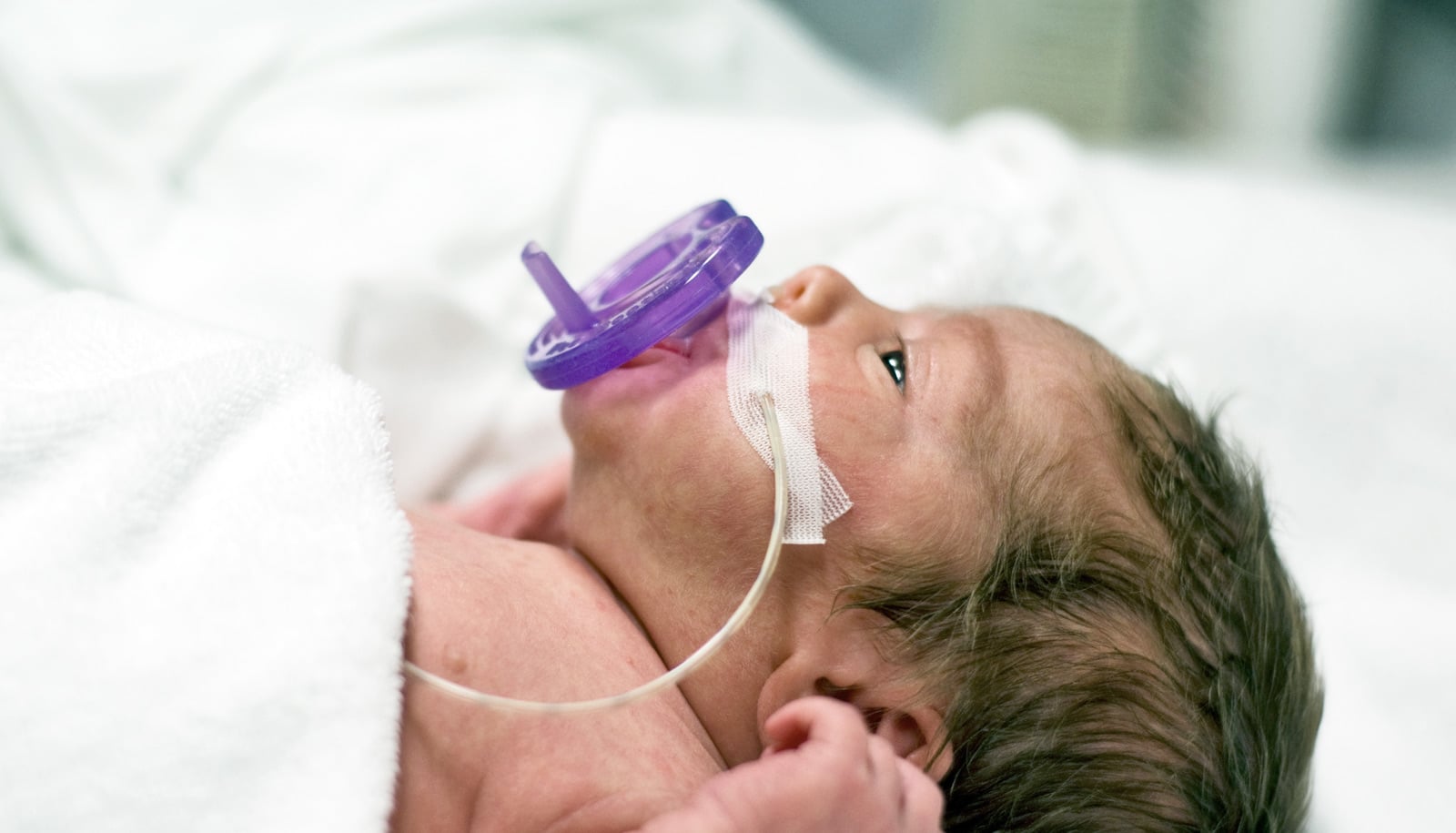
Due to an absence of certain lung cells, mice born into an oxygen-rich environment respond worse to the flu once fully grown. This discovery may explain preterm infants’ added susceptibility to influenza and other lung diseases later in their lives.
The research, published in the American Journal of Respiratory Cell and Molecular Biology, focuses on alveolar type II cells, which help to rebuild lung tissue after damage. When newborn mice are exposed to extra oxygen at birth—which causes their lungs to respond and develop similarly to those of preterm infants—they end up with far fewer of these cells once they reach adulthood.
Once exposed to influenza virus as adults, these mice then developed a much more severe disease than mice born in a traditional oxygen environment.
“We don’t know if this is exactly what happens in preterm infants,” says Michael O’Reilly, professor of pediatrics, environmental medicine, and oncology at the University of Rochester Medical Center.
“But we do know that there’s a direct correlation between the loss of these cells and an inferior response to lung disease, and we do know that there’s something about that early oxygen-rich environment that causes a mouse to respond poorly to viral infection later in life. So this helps connect those dots.”
Artificial placenta could save tiniest premature babies
O’Reilly, who studies the developmental origins of lung disease, hopes to now pursue research on the life cycle of alveolar type II cells. The cells are abundant in the lungs of healthy infants, as they are responsible for producing pulmonary surfactant, a vital compound for the developing lung. As the lungs mature after birth, some of these cells may be pruned away. In theory, the lungs of premature infants take this process too far, pruning too many type II cells.
“Right now, we don’t really understand the biology of that,” O’Reilly says. “But once we do, that opens the door to exploring a potential treatment.”
Source: University of Rochester


