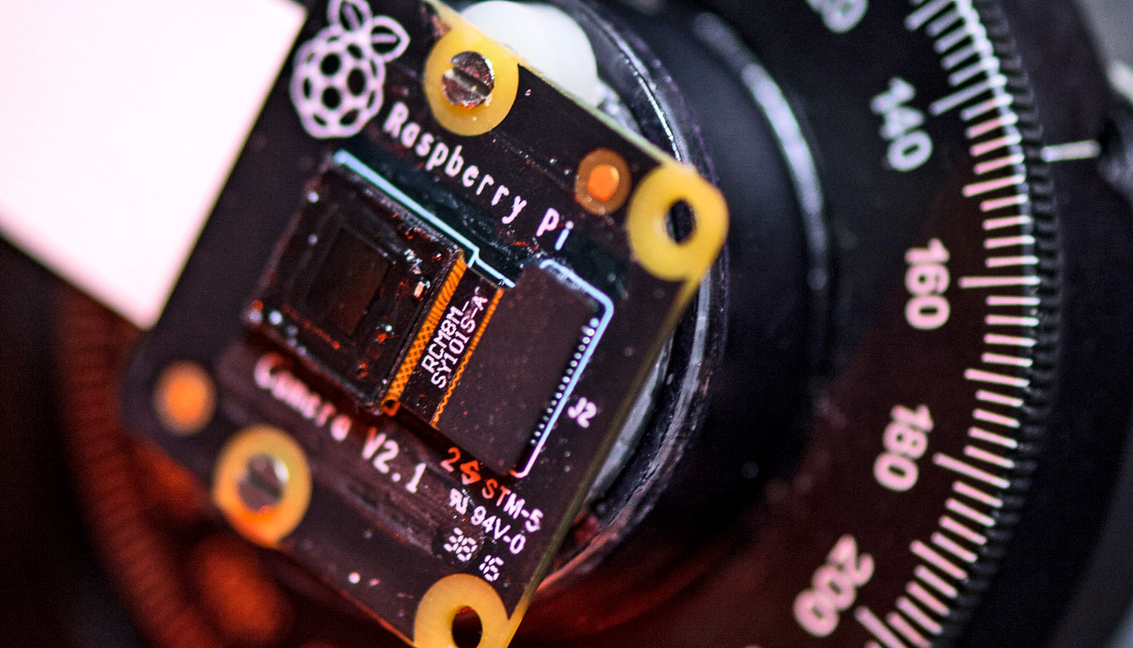
A tiny microscope that weighs only as much as a US penny can capture broad swaths of mouse brain activity with unprecedented resolution, a new study shows.
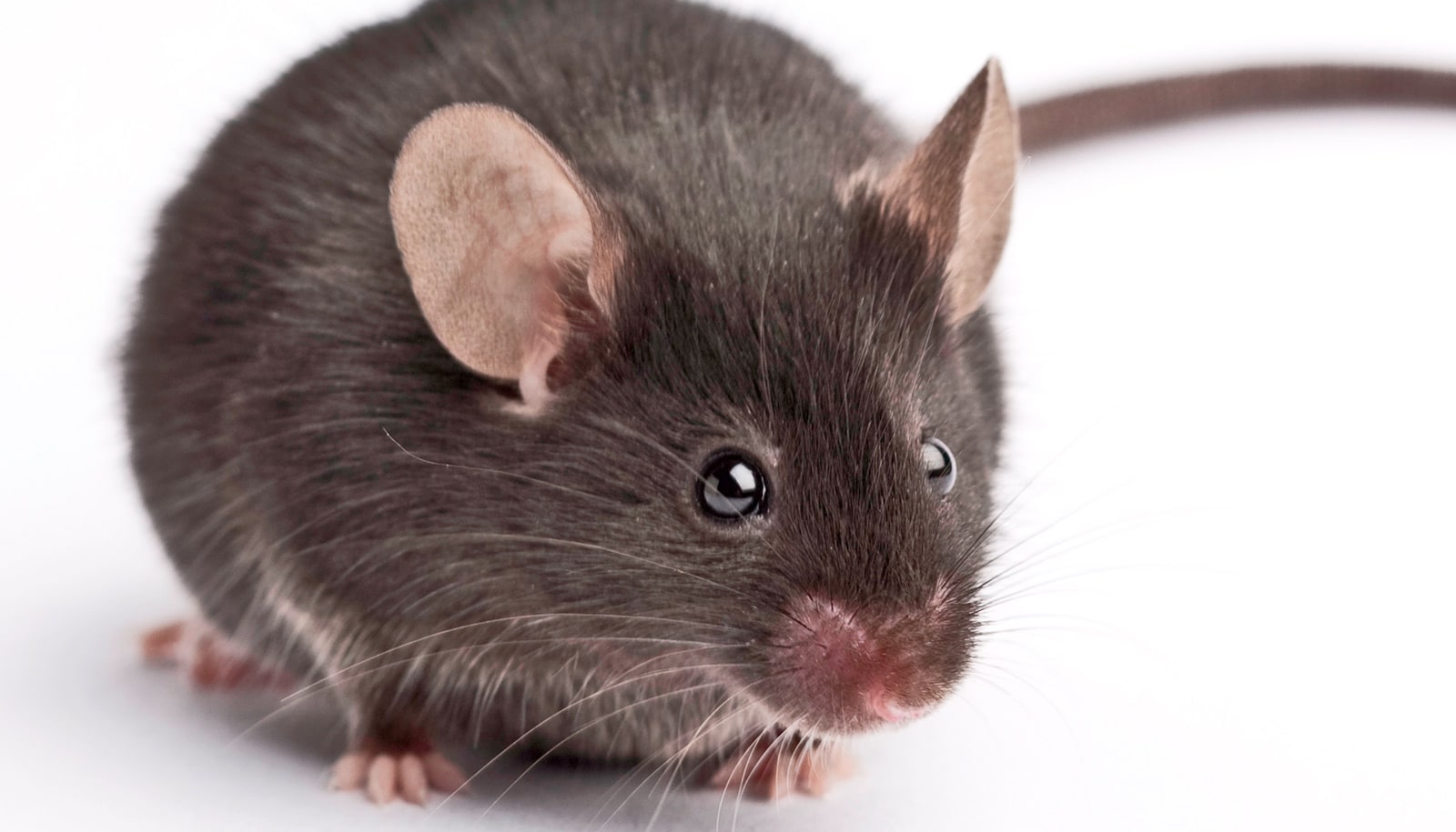
As a mouse explores its environment, millions of neurons across the brain fire in sync. To study only a small subsection at a time would be to miss the forest for the trees, but powerful microscopes capable of capturing the entire mouse brain simultaneously are too heavy to mount on a moving mouse.
“The ability to observe the brain as mice engage in natural behaviors, such as social interactions and prey capture, will advance our understanding of how brain-wide distributed neuroactivity relates to naturalistic behavior,” says Alipasha Vaziri of Rockefeller University who led the study published in Nature Biomedical Engineering.
‘Lens-less’ microscope
Larger mammals can accommodate standard head-mounted microscopes, and even rats can support tech weighing about 20 grams, or eight US pennies. However, mice, which are the go-to model organisms for understanding the brain at work, are much smaller. Microscopes designed to fit them must weigh less than three grams.
“In recent years we’ve seen an explosion of head-mounted microscopes for mice, but they typically support only imaging fields of view of a few hundred micrometers at cellular resolution, since the involved design complexity for larger fields of view comes with an unsustainable weight penalty,” Vaziri says.
Existing models that are light enough for mice to carry invariably compromise the microscope’s field of view, resolution, and depth range (or a combination thereof) and are prone to motion-induced artifacts.
Prior attempts to overcome this limitation were geared toward making whatever technology already existed weigh less—swapping out metal parts for plastic, for instance, while maintaining the basic optical design of microscopes (especially those capable of imaging increased fields of view) in which a heavy lens forms a major part of the weight.
Vaziri addressed this challenge in what he calls “a principled approach.” Instead of trying to make a complex system lens-based system weigh less, he clarified what the technology’s goals really were: solving a high-resolution mapping problem between points in a 3D volume of the sample to points on the 2D surface of a camera. With that in mind, he set out to create a lightweight system that met those goals, without feeling constrained by the need to adapt on an image-preserving lens-based system.
“Everyone was using these multi-element heavy lenses, and trying to make them lighter,” Vaziri says. “Instead of asking how to make lenses lighter, we solved an inverse problem and circumvented the issue, by developing an essentially lens-less strategy and freeing ourselves from the unnecessary constraints of lens-based image formation.”
Lightweight and effective
Enter diffractive optical elements (DOEs). Unlike conventional lenses, which have a continuously curved surface to generate a spherical curvature of the wave front, DOEs use microstructures to manipulate light through diffraction, allowing for precise control of light waves. They are compact, lightweight, and effective.
In microscopy, a traditional lens’s function is to map the points in space on an object onto an image plane (like a camera sensor), ensuring that the image formed resembles the actual scene. However as one tries to form an image of larger and larger field of views while maintaining the resolution, the errors (optical aberrations) caused by a single lens necessitate more lens elements, resulting in a compound lens design.
Using DOEs, the Vaziri lab demonstrated that it is possible to map the positions between the scene and the sensor accurately without forming an image, and then use computational methods to reconstruct the original scene.
Without a hefty compound lens to weigh it down, the mini microscope weighs only 2.5 grams, and provides imaging that can capture broad sections of the mouse brain across a 3.6 x 3.6 mm² field of view with 4 μm lateral resolution, 300 μm depth of field, and recording speed of 16 volumes per second. And most of its parts can be 3D printed, or make use of inexpensive, consumer-grade cell phone camera sensors.
“If labs are interested, they could easily build these microscopes at low cost,” Vaziri says.
Future iterations of the mini microscope may include wireless data transmission—the current model comes with cables that won’t get in the way of a single mouse but could easily get tangled while observing multiple mice interacting with one another—and a fine-tuning of the tech to allow the observation of areas of the brain located deeper within the cortex.
“The system comes with some sacrifices, and is not nearly as high-performance as larger microscopes,” Vaziri says. “But this is a key innovation, and one that could only have come from bringing fresh thinking to the problem and freeing oneself from perceived constraints.”
Source: Rockefeller University