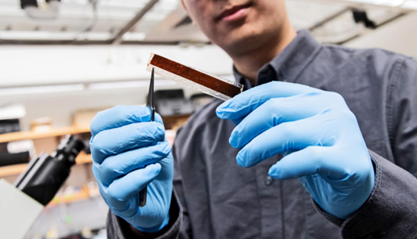
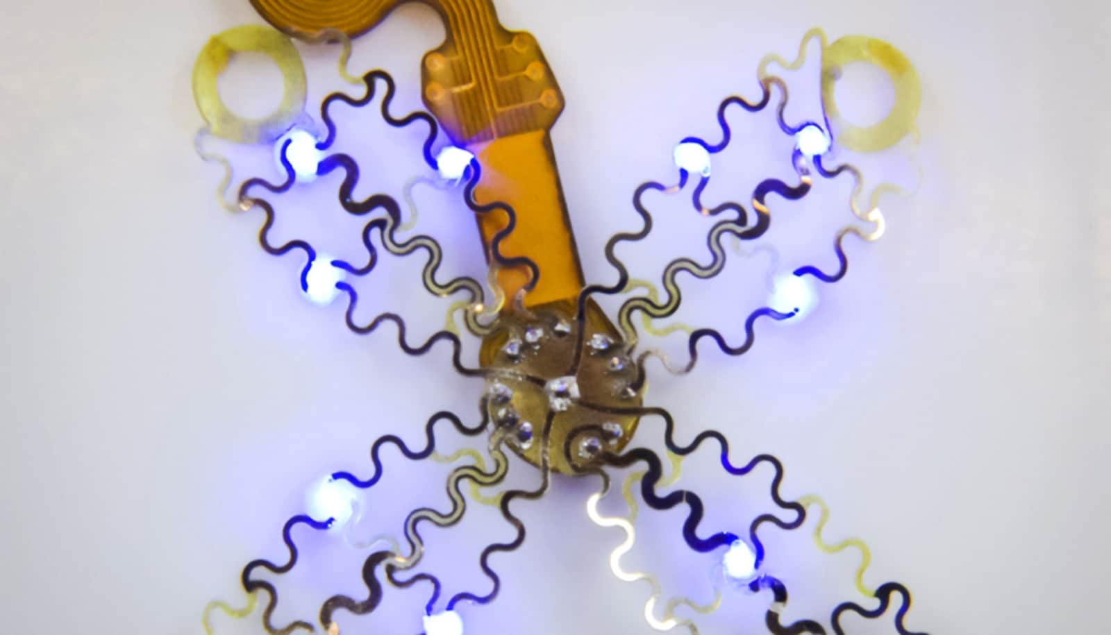
A new wireless pacemaker, powered by light and thinner than a human hair, can be implanted to regulate cardiovascular or neural activity in the body, researchers report.
The featherlight membranes can be inserted with minimally invasive surgery and contain no moving parts.
Millions of Americans rely on pacemakers, small devices that regulate the electrical impulses of the heart in order to keep it beating smoothly. But to reduce complications, researchers would like to make these devices even smaller and less intrusive.
As reported in the journal Nature, the results could help reduce complications in heart surgery and offer new horizons for future devices.
“The early experiments have been very successful, and we’re really hopeful about the future for this translational technology,” says Pengju Li, a graduate student at the University of Chicago Pritzker School of Molecular Engineering and the paper’s first author.
100 times smaller than the finest human hair
The laboratory of Professor Bozhi Tian has been developing devices for years that can use technology similar to solar cells to stimulate the body. Photovoltaics are attractive for this purpose because they do not have moving parts or wires that can break down or become intrusive—especially useful in delicate tissues like the heart. And instead of a battery, researchers simply implant a tiny optic fiber alongside to provide power.
But for the best results, the scientists had to tweak the system to work for biological purposes, rather than how solar cells are usually designed.
“In a solar cell, you want to collect as much sunlight as possible and move that energy along the cell no matter what part of the panel is struck,” Li explains. “But for this application, you want to be able to shine a light at a very localized area and activate only that one area.”
For example, a common heart therapy is known as cardiac resynchronization therapy, where different parts of the heart are brought back into sync with precisely timed charges. In current therapies, that’s achieved with wires, which can have their own complications.
Li and the team set out to create a photovoltaic material that would only activate exactly where the light struck.
The eventual pacemaker design they settled on has two layers of a silicon material known as P-type, which respond to light by creating electrical charge. The top layer has many tiny holes—a condition known as nanoporosity—which boost the electrical performance and concentrate electricity without allowing it to spread.
The result is a minuscule, flexible membrane, which can be inserted into the body via a tiny tube along with an optic fiber—a minimally invasive surgery. The optic fiber lights up in a precise pattern, which the membrane picks up and turns into electrical impulses.
The membrane is just a single micrometer thin—about 100 times smaller than the finest human hair—and a few centimeters square. It weighs less than one fiftieth of a gram; significantly less than current state-of-the-art pacemakers, which weigh at least five grams. “The more lightweight a device is, the more comfortable it typically is for patients,” says Li.
This particular version of the pacemaker device is meant for temporary use. Instead of another invasive surgery to remove the pacemaker, it simply dissolves over time into a nontoxic compound known as silicic acid. However, the researchers say that the devices could be engineered to last to different desired lifespans, depending on how long the heart stimulation is desired.
“This advancement is a game-changer in cardiac resynchronization therapy,” says Narutoshi Hibino, professor of surgery at the University of Chicago Medicine and co-corresponding author of the study. “We’re at the cusp of a new frontier where bioelectronics can seamlessly integrate with the body’s natural functions.”
‘Miraculous achievement’
Though the researchers conducted the first trials with heart tissue, the team says the approach could be used for neuromodulation as well—stimulating nerves in movement disorders like Parkinson’s, for example, or to treat chronic pain or other disorders. Li coined the term “photoelectroceuticals” for the field.
Tian says the day when they first tried the pacemaker in trials with pig hearts, which are very similar to those of humans, remains vivid in his memory. “I remember that day because it worked in the very first trial,” he says. “It’s both a miraculous achievement and a reward for our extensive efforts.”
A screening method Li developed to map the photoelectrochemical output of various silicon-based materials could also have uses elsewhere, Tian points out, such as in fields like new battery technologies, catalysts, or photovoltaic cells.
The research team is currently working with the University of Chicago Polsky Center for Entrepreneurship and Innovation to commercialize the device.
The research made use of the Pritzker Nanofabrication Facility at the Pritzker School of Molecular Engineering and the Electron Microscopy Service of the University of Illinois Chicago Research Resources Center.
The National Institutes of Health, the US Air Force Office of Scientific Research, the National Science Foundation, and the US Army Research Office funded the work.
Source: University of Chicago


