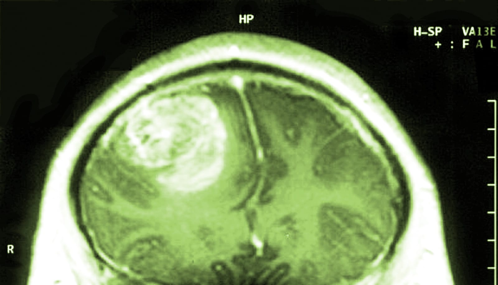
Cancer-hunting stem cells developed from skin cells can track down and deliver a drug to destroy medulloblastoma cells that hide after surgery, according to results from early studies.
Medulloblastoma is the most common brain cancer in children. Previously, researchers showed in preclinical studies they could flip skin cells into stem cells that hunt and deliver cancer-killing drugs to glioblastoma, the deadliest malignant brain tumor in adults.
In the new research, which appears in PLOS ONE, scientists were able to shrink tumors in laboratory models of medulloblastoma and extend life in mice. The study is a necessary step toward developing clinical trials that would see if the approach works for children.
Fewer side effects
The approach holds promise for reducing side effects and helping more children with medulloblastoma, says Shawn Hingtgen, an associate professor in the University of North Carolina’s Eshelman School of Pharmacy, an assistant professor in the neurosurgery department at the university’s School of Medicine, and a member of university’s Lineberger Cancer Center.
More than 70 percent of patients with average-risk disease live five years on standard treatment, but not all patients respond, and treatment can cause lasting neurologic and developmental side effects.
“Children with medulloblastoma receive chemotherapy and radiation, which can be very toxic to the developing brain,” Hingtgen says. “If we could use this strategy to eliminate or reduce the amount of chemotherapy or radiation that patients receive, there could be quality-of-life benefits.”
“The cells are like a FedEx truck that will get you to a particular location, and (deliver) potent cytotoxic agents directly into the tumor.”
Hingtgen and his team showed the natural ability of the stem cells to home in on tumors, and began studying them as a way to deliver drugs to tumors and limit toxicity to the rest of the body. Their technology is an extension of a discovery that won researchers a Nobel Prize in 2012 and showed they could transform skin cells into stem cells.
“The cells are like a FedEx truck that will get you to a particular location, and (deliver) potent cytotoxic agents directly into the tumor,” Hingtgen says. “We essentially turn your skin into something that will crawl to find invasive and infiltrative tumors.”
Shrinking tumors
For the study, researchers reprogrammed skin cells into stem cells, and then genetically engineered them to manufacture a substance that becomes toxic to other cells when exposed to another drug, called a “pro-drug.”
Inserting the drug-carrying stem cells into the brain of laboratory models after surgery decreased the size of tumors by 15 times, and extended median survival in mice by 133 percent. Using human stem cells, they prolonged life of the mice by 123 percent.
The researchers also developed a laboratory model of medulloblastoma to allow them to simulate the current delivery of standard care—surgery followed by drug therapy. Using this model, they discovered that after surgically removing a tumor, the cancer cells that remained grew faster.
“After you resect the tumor, we found it becomes really aggressive,” Hingtgen says. “The cancer that remained grew faster after the tumor was resected.”
There is a need for new treatments for medulloblastomas that have come back, or recurred, as well as for treatments that are less toxic overall, says study coauthor Scott Elton, chief of the UNC School of Medicine pediatric neurosurgery division.
The ability to use a patient’s own cells to directly target the tumor would be “the holy grail” of therapy, Elton says, adding he believes it could hold promise for other rare, and sometimes fatal, brain cancer types that occur in children as well.
“Medulloblastoma is cancer that happens mostly in kids, and while current therapy has changed survival pretty dramatically, it can still be pretty toxic,” he says. “This is a great avenue of exploration, particularly for the 30 percent of children who struggle or don’t make it with standard therapy. We want to nudge the needle even further.”
The UNC Eshelman Institute for Innovation, the UNC Translational and Clinical Science Institute TL1 Pre-doctoral Translational Research Fellowship, and the University Cancer Research Fund supported the work.
Source: UNC-Chapel Hill


