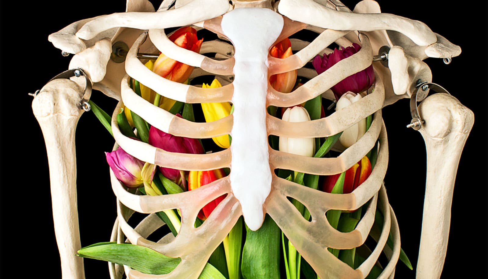
Researchers are succeeding in their efforts to build a better bone graft in the lab.
Each year, about 2.2 million bone-grafting procedures are performed worldwide. The gold standard of care is autografting, which uses the patient’s own bone for tooth implantation and to repair and reconstruct parts of the mouth, face, and skull.
There are drawbacks to autografting that include the need for additional surgery, longer recovery time, complication risks, and the availability of larger amounts of bone.
Having already created a technology that makes bone scaffolds with collagen-like nanostructures, micrometer-sized pores, and natural shapes, the researchers have hit on an “exciting improvement” that regenerates bone by improving cell-matrix interactions, says Peter Ma, a professor of dentistry at the University of Michigan School of Dentistry.
The latest discovery, which is especially beneficial for patients needing repairs involving larger amounts of bone, grew out of a collaboration between the Ma Lab and Franceschi Lab. The team applied for US and international patents of peptide-containing copolymers, nanofiber, and implantable and injectable 3D scaffolds for bone, and other related tissue regeneration that can bring many benefits.
“Having a predictable source of materials to regenerate the bone means much more reliable procedures,” Ma says.
“What is most important is that we can regenerate tissues without introducing exogenous cells, which would potentially complicate the therapies by triggering immune response. The exciting outcome is that our new approach can regenerate about eight times more bone than a scaffold without the special peptides on nanofibers.”
Of the more than 2 million bone graft procedures globally, 500,000 of them are performed in the United States and add up to about $5 billion in costs, the researchers say.
Besides autografting, the team’s new grafting procedure would replace other approaches: allografting, which uses donor tissue, and xenografting, which uses animal tissue. Both can come with risks such as infection and lack of availability.
Ma and colleagues, who describe the science behind the new technique in a study in Bioactive Materials, say there are many benefits to the new approach.
“What we invented are biodegradable polymer templates that contain peptides on nanofibers, acting like keys to open new gates to liberate the locked bone regeneration potential from the recipient’s own cells. After the regeneration of pre-designed 3D bone tissue, the materials will degrade and disappear without potential long-term complications,” says Ma, who is also a professor at the University of Michigan’s College of Engineering and Medical School.
“We are very excited about the discoveries we’ve made. And we believe what we have created can transform bone grafts for the millions of people who require them.”
Source: University of Michigan