Fossils in Brazil indicate a more complex evolutionary history for ray-finned fish brains than previously anticipated, according to new research.
The researchers not only found well-preserved brains in late Paleozoic ray-finned fishes, they also discovered other soft tissues—such as fragments of the heart and eyes, meninges, and gill filaments—a rarity in paleontology due to the scarcity of the fossil record.
“These fossils not only show extensive preservation of soft tissue but also provide a glimpse into the evolution of the brain of fish that lived more than 290 million years ago,” says Rodrigo Tinoco Figueroa, a Brazilian doctoral student at the University of Michigan. “Fossils like this are the only way we can get direct evidence of soft tissue elements from the past. Such information often breaks our expectations about living species.”
Figueroa is the lead author of the new study published in the journal Current Biology. The work is part of his dissertation under paleontologist Matt Friedman in the earth and environmental sciences department.
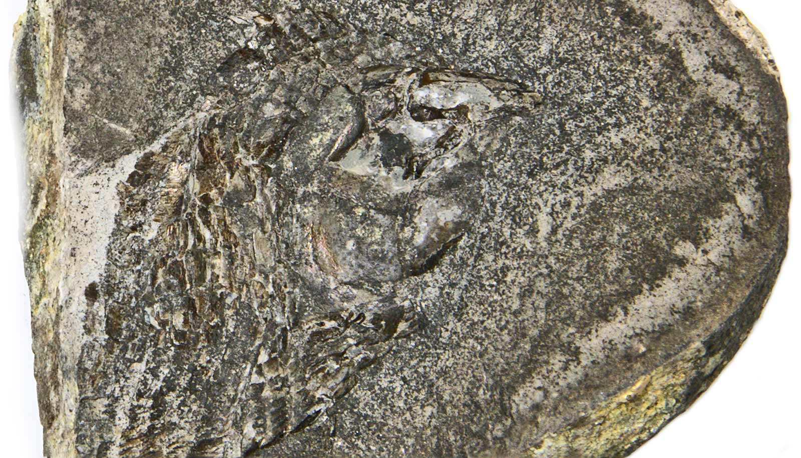
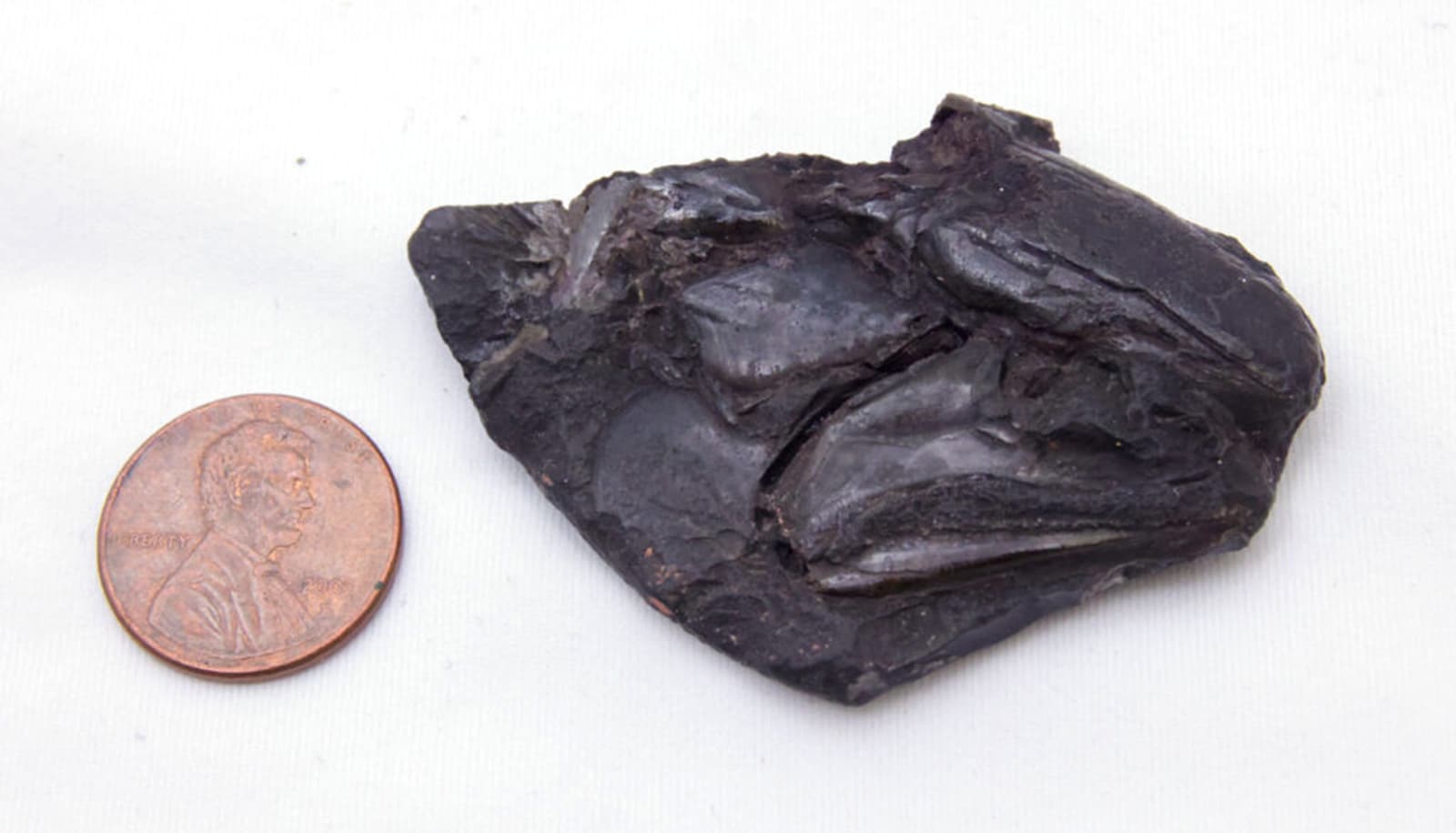
Figueroa says of all the specimens, one called CP 065 is the most surprising.
“In addition to being the first specimen in which I noticed an everted brain, it is also one of the best-preserved fossils I have ever seen,” he says. “Imagine a fossil more than 290 million years old that preserves the brain and its cranial nerves, the delicate meninges that support the brain inside the cranial cavity, gill filaments, fragments of blood vessels, parts of the heart, and possibly skeletal muscles. It is certainly a unique find. Specimens like this are the best way to bring paleontology closer to biology and vice versa.”
Figueroa works with CT scans of fossil ray-finned fish skulls, including these specimens he brought to Michigan on loan from the Paleontological Center of the University of Contestado in Mafra, Santa Catarina, Brazil.
“Using micro-CT of fossils and contrast-enhanced micro-CT of extant species provides us with new three-dimensional data that can go beyond the results provided in this paper, with the addition of new fossils and new comparative material from extant species,” Figueroa says.
In this study, Figueroa scanned eight specimens from Mafra and found some degree of soft tissue fossilization in all of them. In most cases, the brain was preserved in detail, showing morphology similar to that of Coccocephalus, found in previous research.
“After a more detailed examination of all these brains and the associated osteology of the specimens, I was able to determine that there were two distinct species,” he says. “Considering their bone morphology, one seemed closely related to younger fossils, closer to the group that includes all 35,000 living species of ray-finned fish.”
According to Figueroa, these two taxa show different brain morphology.
“This gives us the first evidence of an everted telencephalon in a fossil ray-finned fish,” he says. “This is found in some specimens, while Coccocephalus, from last year’s work, shows the contrasting condition that we call an evaginated telencephalon.”
The researcher says the specimens also preserve detailed evidence of meningeal tissues, such as the membranous tissue supporting the brain inside the head and eyes—including lenses—sclera, muscles, and retinal tissue.
“Although, at the moment, they are not sufficient to provide a clear picture of the evolution of these structures, they are an indication that such extensive preservation of soft tissues is possible,” Figueroa says. “I believe many more discoveries could emerge in the coming years.”
The study is the culmination of five years of research. After discovering the oldest vertebrate fossil brain in 2023, Figueroa wanted to better understand other possible instances of soft tissue preservation in fossils and how much information this type of preservation can provide.
“I still remember the first time I looked at the scan of one of the specimens,” he says. “I was excited just seeing all the details preserved in the bones and then realized there was more. It was an eye in almost pristine condition. From that moment on, it was an adventure to find more and more preserved soft tissue and compare it to living fish. It’s amazing how much these specimens preserve.”
Source: Firnanda Pires for University of Michigan


